 Your new post is loading...
Scar tissue if left untreated/unmanaged could lead to mobility and chronic pain issues. Individuals that are recovering from a traumatic injury often have issues brought on from scar tissue. Scar tissue build-up is part of the healing process but in some cases, the tissue build-up can create another set of health issues. Restricted mobility and range of motion and lack of flexibility can worsen over time. Chiropractors are taught to consider the presence of scar tissue when performing adjustments. This is especially true for patients recovering from a traumatic injury. If left untreated it could affect: - Recovery progress
- Treatment approaches
- The capability of the individual to handle the treatment
- The planning and execution for a chiropractic recovery strategy
Scar Tissue Breakdown Scar tissue can be broken up, managed, and kept loose/relaxed through various chiropractic/physical therapy techniques, stretches, exercises, and diet adjustments. Breaking up scar tissue and keeping it relaxed is necessary to restore full movement and range of motion. A certain degree of scar tissue will remain to mark the wound, but the pliability and softness of these tissues can be treated. Chiropractors can implement several techniques to break down scar tissue. Graston Technique The Graston technique uses instrumentation for addressing scarring in soft-tissue areas like the legs, neck, and lower back. A chiropractor targets the thicker scar tissue areas gently breaking them down. Instrument Adjustment Using a pulsating instrument, chiropractors can target specific areas of scar tissue buildup. The instrument massages the areas to improve flexibility and reduce stress in the tissue. Assisted Manipulation The assisted manipulation technique soothes the area before performing manual adjustments. A chiropractor could use light oil for heat, transcutaneous electrical nerve stimulation to loosen tension, or numbing gel/cream to soften any sensitivity. Trigger point therapy This therapy focuses on heavily scarred tissue areas, where there is substantial buildup. A chiropractor breaks down the scar tissue while continually testing the motion. Therapeutic massage Therapeutic massage is necessary with widespread scar tissue. It improves blood flow and gently stimulates scar tissue to improve movement and alleviate pain. These techniques and how they are applied depends on the individual and the amount of tissue build-up. For example, certain techniques work better for different situations like: - The Graston technique could help after surgery
- Trigger point therapy can help when muscle spasms, and knots present
- Therapeutic massage could be best suited for soft tissue scarring, like whiplash or muscle strains
Adjustment Attention Recent injuries that produce scar tissue can usually be felt while a chiropractor palpates the area, while radiological imaging shows scar tissue from past injuries. A chiropractor will take note of these areas when developing a treatment plan. Severe scar tissue will need special focus, which could mean a longer recovery. This is because the body could take longer to adjust to the adjustments being made. A thorough consultation and investigation will be performed/examined before any adjustments begin. Dr. Alex Jimenez’s Blog Post Disclaimer The scope of our information is limited to chiropractic, musculoskeletal, physical medicines, wellness, and sensitive health issues and/or functional medicine articles, topics, and discussions. We use functional health & wellness protocols to treat and support care for injuries or disorders of the musculoskeletal system. Our posts, topics, subjects, and insights cover clinical matters, issues, and topics that relate and support directly or indirectly our clinical scope of practice.* Our office has made a reasonable attempt to provide supportive citations and has identified the relevant research study or studies supporting our posts. We also make copies of supporting research studies available to the board and or the public upon request. We understand that we cover matters that require an additional explanation as to how it may assist in a particular care plan or treatment protocol; therefore, to further discuss the subject matter above, please feel free to ask Dr. Alex Jimenez or contact us at 915-850-0900. The provider(s) Licensed in Texas& New Mexico* References Shin, Thuzar M, and Jeremy S Bordeaux. “The role of massage in scar management: a literature review.” Dermatologic surgery: official publication for American Society for Dermatologic Surgery [et al.] vol. 38,3 (2012): 414-23. doi:10.1111/j.1524-4725.2011.02201.x
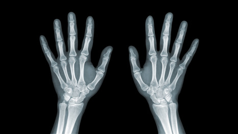
Wrist & Hand Trauma - Distal Radius & Ulnar Fractures (Colles, Smith's, Barton's, Chauffeur's, DiePunch)- complicated by 50% ulnar styloid Fx, TFC path, DRUJ dislocation, scapholunate lig dissociation, lunate/perilunate dislocation )
- Carpal bones Fracture & dislocations (scaphoid, triquetrum, hamate Fx &Lunate/perilunate dislocation)
- Ligaments dissociation (Scapholunate dissociation, Lunotriquetral instability)
- Metacarpal & Phalangeal fractures (Bennett, Rolando, Game keeperFx/Stener lesion, Boxer Fx)
- Pediatric wrist injury (green-stick Fx, Torus Fx, Bowing/plastic deformity, Salter-Harris injuries)
- In all cases, Orthopedic hand surgical referral is required
- Colles fx: m/c d/t FOOSH+pronation. m/c inOSP/elder women. Rare in men and if occurs need DEXA to avoid hip Fx etc. Young pts: high-energy trauma. Typically extra-articular.50%-cases show Ulna styloid (US) Fx.
- Complications: dinner fork deform, CRPS, DJD, nerve entrapment.
- Imaging: x-rad is sufficient, CT in complex Fx, MRI helps with ligament tears and TFC.
- Rx: if extra-articular and <5-mm distal radius shortening and <5-degree dorsal angulation closed reduction+casting is sufficient. ORIF in complex cases.
- Image Dx: distal rad impaction/shortening,dorsal angulation of distal fragment, carefully examine if intra-articular extension, 50% US Fx
- Smith Fx: Goyrand in French literature. Considered as reversed Colles, otherwise almost identical, I.e., 85% extra-articular, 50%US Fx, OSP/elderly women, young pts-high-energy trauma. Differences: mechanismFOOSHwith flexed wrist thus m. Less frequent.
- Imaging steps: (see Colles Fx) C
- Complications: similar to Colles Fx
- Rad Dx: 85% extra-articular with volar(anterior) angulation of the distal fragment,radial shortening. Carefully examine cortical breach suspecting intra-articular extension that can be named as Smith type 2 or Reversed Barton Fx (next)
- Rx: similar approach as in Colles.
- Barton fx: FOOSH, impaction of distal radius similar to Colles but the Fx line extends from the dorsal radial aspect into radiocarpal joint resulting with dorsal slip/dislocation of the carpus.
- Imaging: 1st sept x-radiography often with CTto examine intra-articular Fx extension and operative planning
- Rad Dx: distal radius Fx extending from dorsal into the radiocarpal joint with a variable degree of displacement, the proximal slip of the carpus
- If Fx line extends from the volar aspect into the wrist joint named Reversed Barton aka Smith type 2 (above bottom image)
- Complications: similar to all distal radius Fx
- Rx: operative with ORIF
- Chauffeur's/backfire Fx aka Hutchinson Fx: intra-articular Fx of Radial styloid. The name derives from the time when the car had to be started with a hand crank that could backfire inducing wrist dorsiflexion and radial deviation.
- Imaging: x-radiography is sufficient. CT may be helpful if Fx not readily shown by x-rays.
- Complications: non-union, malunion, DJD,scapholunate dissociation,lunate/perilunate dislocation
- Rx: operative with percutaneous lagscrewin all cases d/t intra-articularnature
- Die-Punch Fx: impaction Fx by the Lunate bone into distal articularLunate fossa of the Radius. IntraarticularFx. Derives its name from a technique to shape (impress) a hole in industrial machining "die-punch."FOOSH injury.
- Imaging: 1st step x-rays, may be equivocal d/t subtle depression of the Lunate fossa then CT scanning is most informative.
- Rad Dx: impacted lunate fossa region with intra-articular Fx extension. This can present as a comminuted Fxarticular Fx of the Distal Radius.
- Rx: operative d/t intra-articular Fx
Construct arcs of Gilula when evaluating carpal injuries. An Important step required to avoid missing subtle changes in carpal alignment and cortical continuity - Scaphoid bone Fx: m/c Fx carpal bone. D/tFOOSH wrist extended radially deviated. Location of Fx is most important to prognosis: Waist-m/c location (70%). May have 70-100%chance of AVN. Proximal pole Fx: 20-30% with a high risk of non-union. Distal pole-10%shows better prognosis. Distal pole Fx is m/c in children. Key clinical sign; pain in the snuffbox.
- Imaging: 1st step-x-radiography but 15-20%missed d/t occult Fx. Special views required. Thus MRI is the most sensitive and specific for early occult Fx. Bone scintigraphy has98/100% specificity & sensitivity esp. 2-3 days after the onset. Key rad. Dx: Fx line if evident, displacement and obscuration of scaphoid(navicular) fat pad, examine for scapholunate dissociation. If proximal bone appears sclerotic-AVN occurred. MRI: low on T1 & high on T2/STIR/FSPD d/t bone edema, a low signalFx line can be noted.
- Rx: Spica cast should be applied if clinically suspected even w/o x-ray findings. For waistFx-cast for 3-mo for prox pole 5-mo immobilization. ORIF or percutaneous pinning with a Herbert screw.
Scapholunate Ligaments Dissociation - SNAC wrist: scaphoid non-union advanced collapse. Often d/t non-union and dissociation of scapholunate ligaments (SLL)with progressive radiocarpal and intercarpalDJD. The Proximal scaphoid fragment is attached to Lunate with distal dissociating and rotating‘signet ring” sign on x-rays.
- SNAC wrist may often result in DISI
- Rx: progressive DJD may lead to four-corner arthrodesis
- Scapholunate advanced collapse (SLAC wrist): SLLdissociation with progressive intercarpal and radiocarpal DJD and volar or dorsal carpal displacement (DISI & VISI). Causes: trauma, CPPD, DJD, Kienboch disease (AVN of Lunate), Preiserdisease (AVN of Scaphoid).
- SLL dissociation will lead to Dorsal or VolarIntercarlate aka Intercarpal Segmental Instability (DISIor VISI).
- Rad Dx: Dx underlying cause. X-rays demonstrate dorsal or volar angulation of the Lunate with increased or decreased scapholunate angle on the lateral view. On frontal view: Terry Thomas sign or widening of scapholunate distance 3-4-mm as the upper limit of normal.
- MRI may help with ligament evaluation and pre-surgical planning
- Rx: often operative with late DJD. Four-corner arthrodesis
- Triquetrum Fx: 2nd m/c carpal bone Fx. M/C dorsal aspect is avulsed by the tough Dorsal radiocarpal ligament. Cause: FOOSH.
- Imaging: x-radiography wrist series is sufficient. Best revealed on the lateral view as an avulsed bone fragment adjacent to the dorsum of the Triquetrum. CT may help if radiographically equivocal.
- Rx: conservative care
- Complications: rare, may persist as pain on the dorsum of the wrist
- Hook of the Hamate Fx: m/c occurs in batting sports (cricket, baseball, hockey, impact by a golf club, etc.) 2% of carpusFx.
- Imaging: x-radiography may fail to detect an Fx unless "carpal tunnel view" is used. CT may help if x-rays unrewarding.
- Clinically: pain, positive pull test, weak, painful grip. Deep ulnar n. Branch may be affected by the Guyon canal.
- Rx: usually non-operative, but chronic non-union may require excision.
- DDx: bipartite hamate
- Lunate vs. Perilunate dislocation: Lunate is m/c dislocated carpal bone. Overall infrequent carpal injury. However, often missed!
- Occurs with FOOSH and wrist extended and ulnar deviated. Imaging: 1st step x-rays. Ifunrewarding or require more complex injury evaluation CT scanning.
- Key Rad DDx: DDx Lunate from perilunate dislocation. Lunate dislocation: lunate lost its contact with distal radius ‘spilled teacup” on the lateral. Perilunate dislocation: Lunate maintains its contact with distal radius despite the Capitatedorsally dislocated. Lunate dislocation is additionally helped to identify a “pie sign” d/t Lunate overlapping the Capitate
- Rx: emergency reduction and operative repair of torn ligaments
Metacarpal & Phalangeal Injuries - Bennett Fx: intra-articular but noncomminuted impact-type Fx of the base of 1st MC bone of the thumb. X-radiography is sufficient.
- Rad Dx: characteristic triangular fragment of bone on the ulnar aspect of the 1st MCbase, often with radial subluxation of the remaining radial aspect of the 1st MC
- Complications: DJD, non-union, etc.
- Rx: prone to instability/non-union requiring an operative care
- Rolando Fx: aka comminuted Bennett with Y or T-configuration. More complex injury. It is unstable requiring operative care
- Gamekeeper thumb: traditionally described as a chronic tear of the ulnar (medial) collateral ligament at 1stMCP in English Gamekeepers' who performed neck twisting/killing of small game. An acute injury may also be named as Skier's thumb. This injury can be ligamentous w/o a fracture and/or an avulsion injury at the 1st proximal phalanx base.
- Complication: Stener lesion or displacement of torn ligament over Adductor pollicis muscle that cannot heal w/o surgical repair. MRI Dx is required.
- Avoid thumb stress views that can induce a Stenerlesion
- Imaging: x-radiography followed by MRI to Dx Stenerlesion. MSK US can be used if MRI is unavailable.
- Stener lesion on MRI & MSUS: ulnar collateral stump is more superficial to Adductor pollicis aponeurosis and appears like a low signal mass-like stump forming so-called "yo-yo on the string sign" reported both on MRI and MSK US.
- Rx: often operative
- Boxer Fx: m/c MC Fx. An extra-articular usually non-comminuted or minimal comminuted Fx through m/c the 5th and sometimes the 4th MCneck-head junction (occasionally through the shaft) resulting in volar head angulation. Mechanism: direct impact as in clenched fist punching hard surface (e.g., facial bones/wall punching) hence 95% in young males.
- Imaging: x-radiography hand series is sufficient
- Rad Dx: Fx line transverse or oblique through MCneck with volar head angulation. Evaluate the degree of displacement, critical to report.
- Rx: typically non-operative with short –arm gutter splint and digits flexed. (https://www.aafp.org/afp/2009/0101/p16.html)
- N.B. If the same mechanism fractures the 2ndand 3d MC in the same anatomic area, it may require operative care.
- Phalangeal hand Fx: m/c skeleton Fx (10% of all Fx). Sports and industrial injuries dominate
- Imaging: x-radiography with hand series or PA/lateral finger views will suffice
- Rad Dx: if prox phalanx Fx, distal fragment is angled volarly with prox fragment dorsally. Distal phalanx may be angled dorsally. Key observation: nail bed injury, which considered an open Fx with a risk of infection.
- Rx: if <10-degree angulation-buddy-taping with motion rehab. CRPP vs. ORIF can be considered in complex cases-Orthopedic hand surgeon referral
- Complication: loss of motion, necrosis, infection.May result with amputation
- For additional common injuries: PIP is m/c dislocated joint. Mullet (Baseball) finger, Jersey finger and other injury refer to:
- https://www.aafp.org/afp/2012/0415/p805.html
- Felon: septic infection of the fingertip pulp typically with Staph.Aureus. Causes: needle prick(diabetics), paronychia, nail splinters, etc. m/c in index and thumb, presenting with pain, swelling, etc.
- D/t specific pulp anatomy theinfection>swelling leads to pulp compartment syndrome-pressure and necrosis.
- Rx: operative with incision distal to DIP, irrigation/debridement
Pediatric Wrist Injury - Incomplete Fx: Greenstick Fx, Torus (Buckle)Fx, Bowing (Plastic) deformity/Fx. D/t FOSHe.g. fell off the monkey bar. m/c affects <10-years-old.
- Key Imaging diagnosis: degree of angulation/displacement, epiphyseal growth plate injury (Salter-Harris classification)
- Rx: usually non-operative (closed reduction and casting)
- Distal Radioulnar Joint (DRUJ) Instability-common injury following trauma as in FOOSHwith wrist hyperextension and rotation and disruption of DRUJ ligaments and TFCcomplex. Avulsion of ulnar styloid with the dorsal or volar displacement of distal ulnar should be noted.
- Imaging steps: x-rays initially, MRI may identify ligaments and TFC damage, MSKUScan help with ligaments tearing.
- Note: isolated DRUJ volar (top image) and dorsal (bottom image) dislocation.
Wrist & Hand Arthritis - Wrist DJD-typically is secondary to trauma, scapholunate dissociation, SLAC, SNAC wrist, CPPD, Keinboch or Preiser Disease and others.
- May lead to major functional loss
- Imaging: typically presents as radiocarpal JSL, subchondral sclerosis,osteophytosis, subcortical cysts, and loose bodies. Typically additional induces intercarpal degeneration and particularly Tri-scaphe joint.
- MRI may be helpful with early recognition of scapholunate dissociation, Lunate/Navicular AVN.
- Rx: conservative vs. operative.
- DJD Hand: Extremely common. True primary OA. MCP-never affected w/o DIP & PIP
- If isolated MCP OA noted considerCPPD & Hemochromatosis (Hook-like osteophytes)
- Clinically:
- Mid-age females
- Typically painless except 1st CMC OA
- DIPs-Heberden nodes, PIPs-Bouchard nodes
- Erosive OA (occasionally called“inflammatory OA”)
- A Spectrum of OA but producing central proximal erosions at DIPs and PIPsresulting with very characteristic “gullwing” appearance. No systemic inflammation (no CRP, RF, Anti-CCP Ab)typically in middle-aged/elderly females, like Hand OA, often seen in families
Rheumatoid Arthritis - Rheumatoid Arthritis (RA)-chronic systemic inflammatory disease of unknown etiology, targeting synovial joints, tendons with multiple systemic involvement (lung, CVS, Ocular, Skin, etc.) Pathology: Tcell>Macrophage/APC>mediatedautoimmune process resulting in pannus formation and gradual destruction of ST, cartilage, bone, and other tissues. 3% FemalesVS.1% Males. Environmental triggers: infection, trauma, smoking, and others in a genetically susceptible individual. 20-30%may be disabled after 10-years.
- Dx: clinical, labs, imaging.Symmetrical Polyarthritis esp. in MCP, wrists (2nd & 3RD MCP)
Meniscal Tears - Acute or chronic. Imaged with MRI (95% sensitivity & 81% specificity)
- Menisci are formed by a composition of radial and circumferential collagen fibers (97% type 1) mixed with cartilage, proteoglycans, etc. 65-75% H2O
- Aging can lead to meniscal attrition
- Acute tears are d/t rotational and compressive forces, ACL deficient knees show greater chances of meniscal tears
- Posterior horn of medial meniscus is m/c torn except in acute ACL tears when the lateral meniscus is m/c torn
- The meniscus is well vascularized in children. In adults, 3-zones exist: inner, middle and outer (above bottom image)
- Injury of the inner zone has no chance of healing
- Injury of the outer zone (25% in total) has some healing/repair
Clinical Presentation - Pain, locking, swelling
- Most sensitive physical sign: pain on palpation at the joint line
- Tests: McMurry, Thessaly, Apply compression in prone
- Management: conservative vs. operative depends on location, stability, patient's age, and DJD and the type of tear
- Partial meniscectomy is performed. 80% proper functions on follow up. Less favorable if >40-y.o and DJD
- Total meniscectomy is not performed and only viewed historically. 70% OA 3-years after surgery 100% OA after 20 years post surgery.
Axial MR - Appearance the medial (blue) and the lateral meniscus (red)
Menisci Play Significant Role Types Location & Stability - Types, location, and stability of tears are v. important during MRI Dx
- Vertical/longitudinal tears especially occur in acute ACL tears. Some longitudinal tears found at the periphery or "red zone" may heal
- Bucket handle tear: longitudinal tear in the inner edge that is deep and vertical extending through the long axis and may displace into a notch
- Oblique/flap/parrot-beak are complex tears
- Radial tear at 90-degree to plateau
Axial T2 - Axial T2 WI fat-sat and coronal STIR slices of the posterior horn of the medial meniscus.
- Note a radial tear of the posterior horn of the medial meniscus near the meniscal root. This is potentially an unstable lesion requiring operative care
- The meniscus, in this case, is unable to provide a "hoop-stress mechanism."
MRI Slices Coronal & Sagittal - Fat-sat coronal and sagittal proton density MRI slices revealing horizontal (cleavage) tear that is more typical in the aged meniscus
- In some cases, when this tear does not contain a radial component, it may partially heal obviating the need for operative care
T2 w GRE Sagittal MRI Slice - Complex tear with a horizontal oblique and radial component.
- This type of tear is very unstable and in most cases may need operative care
Bucket Handle Tear - Bucket handle tear are m/c in the medial meniscus esp. with acute ACL and MCL tear
- MRI signs; double PCL sign on sagittal slices
- Absent "bow-tie" sign and others
- Most cases require operative care
DDx From Meniscal Degeneration - Occasionally meniscal tears need to be DDx from meniscal degeneration which may also appear bright (high signal) on fluid-sensitive MRI
- The simplest rule is that if there is a true meniscal tear aka Grade 3 lesion, it always reaches/extends to the tibial plateau surface
The Role of MSK Ultrasound (US) in Knee Examination - MSK US of the knee permits high resolution and dynamic imaging of primarily superficial anatomy (tendons, bursae, capsular ligaments)
- MSK US cannot adequately evaluate cruciate ligaments and the menisci in their entirety
- Thus MR imaging remains modality of choice
Potential Pathologies Successfully Evaluated by MSK US - Patellar tendionosis/patellar tendon rupture
- Quadriceps tendon tear
- Prepatellar bursitis
- Infrapatellar bursitis
- Pes Anserine bursitis
- Popliteal cyst (Baker cyst)
- Inflammation/joint effusion with synovial thickening and hyperemia can be imaged with US (e.g., RA) especially with the addition of color power Doppler
Patient Presented With Atraumatic Knee Pain & Swelling - Radiography revealed sizeable soft tissue density within the superficial pre-patella region along with mild-to-moderate OA
- MSK US demonstrated large septated heterogeneous fluid collection with mild positive Doppler activity on the periphery indicating inflammation d/t Dx of Superficial pre-patella bursitis
Long Axis US Images - Note normal lateral meniscus and fibers of LCL (above bottom image) compared to
- Horizontal degenerative cleavage tear along with protrusion of lateral meniscus and LCL bulging (above top image)
- Major limitation: unable to visualize the entire meniscus and the ACL/PCL
- MRI referral is suggested
Rupture of Distal Tendon of Quadriceps - Note rupture of distal tendon of the Quadriceps muscle presented as fiber separation and fluid (hypo to anechoic) fluid collection within the substance of the tendon
- Advantages of MSK US over MRI to evaluate superficial structures:
- Dynamic imaging
- Availability
- Cost-effective
- Patient's preparation
- Disadvantages: limited depth of structures, inability to evaluated bone and cartilage, etc.
Osteochondral Knee Injuries (OI) - osteochondral knee injuries can occur in children 10-15 y.o presented as Osteochondritis Dissecance (OCD) and in mature skeleton m/c following hyperextension and rotation trauma, particularly in ACL tear.
- OCD-typically develops from repeated forces in immature bone and affects m/c postero-lateral portion of the medial femoral condyle.
- OI in mature bone occurs m/c during ACL tears mainly affecting so-called terminal sulcus of the lateral femoral condyle at the junction of the weight-bearing portion opposed to the tibial plateau and the part articulating with the patella
- Osteochondral injuries may potentially damage the articular cartilage causing secondary OA. Thus need to be evaluated surgically
- Imaging plays an important role and should begin with radiography often followed by MR imaging and orthopedic referral.
OCD Knee - 95% associated with some trauma. Other etiology: ischemic bone necrosis especially in adults
- Other common location for osteochondral injuries: elbow (capitellum), talus
- 1st step: radiography may detect osteochondral fragment potentially attached or detached
- Location: the posterior-lateral aspect of the medial femoral condyle. Tunnel (intercondylar notch) view is crucial
- MRI: modality of choice >90% specificity and sensitivity. Crucial for further management. T1-low signal demarcating line with T2 high signal demarcating line that signifies detachment and unlikely healing. Refer to orthopedic surgeon
- Management: stable lesion esp. in younger children>off weight-bearing-heals in 50-75%
- Unstable lesion and older child or impending physeal closure>operative fixation.
Hip Fractures - Garden Classification (above) helps with Dx and correct management of patients
- M/C Fx are subcapital (80%)
- Fxs differentiated as intra-capsular (high risk of AVN) & extra-capsular (lower risk of AVN)
- Garden 1: incomplete undisplaced Fx typically impaction with valgus off-set of the head (15-20% AVN) patient able to ambulate
- Garden 2: complete, undisplaced Fx (30% AVN)
- Garden 3: complete, partially <50% displaced
- Garden 4: complete, 100% displaced Fx, pt collapsed with entire LE in ER (below image)
- Most osteoporotic Fx are intracapsular
Complete Displaced Femoral Neck Fracture Clinical Presentation Imaging: Begins with X-radiography with Most Fxs - CT scanning may help with further delineation of Fx complexity/displacement and Dx of additional regional Fxs
- MRI can be helpful if x-radiography fails to Dx fx
- X-radiography pitfalls: some undisplaced Garden 1 & 2 Fxs may be missed d/t pre-existing DJD and osteophytes along the femoral head-neck junction that may overly the Fx line
- Fx line is incomplete and too small/subtle especially if the study is read by non-radiologists
- Incomplete Fxs if left untreated will not heal and likely to progress to complete Fxs
- AP hip spot view: note valgus deformity of the head (above yellow arrow) with a small/subtle line of sclerosis in the sub-capital region representing Garden 1 Fx. MRI may help with Dx of subtle radiographic Fxs. If MRI contraindicated, Tc 99 radionuclide bone scan may help demonstrate high uptake of the radiopharmaceutical in Fx (below image)
Above - Tc99 Radionuclide Bone Scan Reveals Left Subcapital Femoral Neck Fx - Garden 2 complete undisplaced (above green arrows) Fx
- AP hip: Garden 3 complete partially displaced Fx (above the first image)
- AP pelvis: complete displaced Garden 4 Fx (above the second image)
- Clinical pearls: in some cases of Garden 4 Fx, DDx may be difficult to differentiate from OSP vs. pathologic fx d/t to bone Mets of Multiple myeloma (MM)
- Management: depends on patients age and activity level
- Garden 3 & 4 require total hip arthroplasty in patients <85-y.o.
- Garden 1 & 2 may be treated with closed reduction of fx and open capsule and 3-cannulated fixating screws
- Pre-existing DJD may require total arthroplasty
- Occasionally observation may be performed on patients who are not active and significant risks of surgery and depends on surgical centers
- m/c Rx of Garden 1 & 2 undisplaced Fx with 3-screws. Screws proximity depends on the bone quality and Fx type
- THA aka hip replacement: cemented THA with bone cement (above the first image) vs. non-cemented (biologic) that is used mostly in younger patients
- 2-types: metal on metal vs. metal on polyethylene
- The femoral angle of the prosthesis should have slight valgus but never >140 degrees
- The non-cemented component uses porous metal allowing the bone to integrate sometimes coating in bone cement from osteoconduction
- THA has good outcome and prognosis
- Occasionally cement failure, fractures, and infections may complicate this procedure
Supplemental Reading
|
Spinal Cord Injuries Are Not Only Caused by Trauma When people think of spinal cord injury/s or SCI's, traumatic events like a major auto accident, hard fall, severe sports injury or intense work injury come to mind. It is true that auto accidents are the leading cause, however, non-traumatic accidents and diseases like a spinal tumor can also cause spinal cord injuries. These types of injuries involve damage to the spinal cord that can temporarily or permanently affect its functionality. Spinal cord injuries are divided into 2 categories: traumatic and non-traumatic. Even with non-traumatic injuries the impact or severity is not lessened and the aftermath can have a devastating effect on a person’s life. Spinal Cord Injury Trauma - Vehicle crashes: Motor vehicle accidents are the leading cause of traumatic spinal cord injuries, and they account for 40% of all SCI's.
- Falls: Falls are the second cause of traumatic spinal cord injuries, and they account for 32% of injuries.
- Sports: Sports and recreational activities cause around 9.0% of traumatic spinal cord injuries.
- Violence: Violent acts, like gunshot wounds or a stabbing cause around 14% of spinal cord injuries.
Spinal cord injuries occur more in men than women with 80% of cases affecting men. Although people of all ages can experience SCI, there are activities/events that increase the risk that affects different age groups more than others. An example is high-impact actions like motor vehicle accidents and sports injuries occur more in young adults. Conversely, spinal cord injuries caused by a fall happens more in adults over age 60. Regardless, SCI occurs more in the neck or cervical spine. Around 60% of cases involve the neck, followed by the mid-back or thoracic spine that averages to around 32% of injuries. Although most people experience low-back pain, only 9% of spinal injuries occur in the low back and tailbone or lumbosacral spine. Understanding SCI Trauma Damage to the spinal cord not only affects the area where the point of impact occurred. The primary injury can also damage cells, dislocate the vertebrae and cause spinal compression. It can also trigger secondary injuries, that cause a series of biological changes. This can happen within weeks or months after the injury. The secondary injury cascade processes: Glial cells along with the nerve cells in the spinal cord begin to die. These cells provide nutrients and other support to the nerve cells in the central nervous system. This consists of the brain and spinal cord. The blood vessels in the spinal cord lose their ability to function, and this reduces the blood supply to the cord. When the blood supply becomes inadequate it is called ischemia. Blood vessel injuries expose the cord to inflammatory cells that in turn cause swelling. When the spinal cord becomes inflamed the spinal cord gets more and more compressed. If this happens the initial injury progressively worsens. This changes the cord’s structure and its normal operation. The secondary injury cascade can interfere with the cord’s ability to heal itself. This means an individual could experience permanent nerve pain and dysfunction. Non-traumatic SCI Spinal cord damage does not only happen with traumatic events. An SCI can also be caused by non-traumatic diseases in the spine. Tumors are the leading cause, but infections and degenerative disc disease can also cause serious damage. Non-traumatic SCI's happen more than traumatic based ones. The incidence of traumatic SCI in North America comes to around 54 cases per one million people. With the incidence of non-traumatic SCI is around 1,227 cases per million people. Healthy Spinal Cord Injury Outcomes Spine researchers are making great strides in developing optimal protective and regenerative treatments to improve spinal cord health after these injuries occur. Currently, innovative medical, surgical, cell-based and alternative treatments are furthering the medical community’s understanding of SCI's. This is dramatically improving the quality of life and creating positive futures for individuals who experience these injuries. NCBI Resources The muscles in the back keep the spine moving and functioning properly. When the spine or abdominal muscles are weak this creates a higher probability of a back strain or injury. Having strong, healthy spine muscles are important because they function in maintaining correct posture, which in some cases, causes chronic back pain because of poor posture. If only one part of the body is strengthened like the back is not enough. Therefore strengthening the rest of the body is a must. These include the body’s core and leg muscles. Total body strength will reduce back pain and can help perform regular activities, like lifting heavy objects much easier, with more confidence and with a lesser probability of injury.
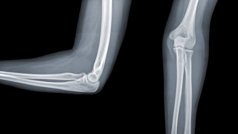
Acute Elbow Trauma - In adults: Radial head Fx is the m/c (33%) and accounts for 1.5-4% of all fractures. Etiology: FOOSH with forearm pronated. Associated injuries: elbow collateral ligaments tears. EssexLoprestiFx with interosseous membrane tearing and dislocation of the Distal Radio-Ulnar Joint(DRUJ)
- Terrible triad: of the Radial head Fx, elbow dislocation and Coronoid process Fx (typically avulsed by the Brachialis M)
- Imaging: 1st step is x-radiography with elbow series, CT scanning may help in complex cases, MRIif ligamentous injury.
- In children: Supracondylar Fx of the distal humerus accounts for 90% of acute trauma. It is always d/t accidental trauma with FOOSH and elbow extended, rarely <5% with flexed elbow. MostSupracondylar Fx occur in children <10 y.o. Males>Females. Complications: malunion in cubitus varus aka Gunstock deformity, vascular injury and acute ischemic compartment syndrome with Volkmann contracture
- Imaging: 1st step x-radiography can be sufficient. CT occasionally used in complex cases.
- Radial head (RH) Fx: Mason classification helps to determine the degree of complexity and mode of treatment
- Type 1- undisplaced is the m/c and stable contained by ligaments. On radiographs can be very subtle and evaluation of abnormal elbow fat pads is critical and often the only diagnostic clue
- Type 2- displaced by 2-mm or > with rotational block
- Type 3- comminuted >2-3 fragments and
- Type4 is presented with RH fx, posterior elbow dislocation and sometimes Coronoid process fracture often d/t Brachialis M avulsion
- Rx: Type 1 managed non-operatively by immobilization and movement rehab. Type 2- ORIF if rotational block. Type 3 and 4, ORIF and RH resection or RH arthroplasty
- Note abnormally displaced anterior fat pad (orange arrow) and the emergence of the posterior fat pad (green arrow) that is usually deep in the olecranon fossa and not seen unless acute hemarthrosis or other effusiondevelopsFat pad signs are most reliable indicators of intra-articular elbow Fx
- Mason type 1 RH Fx can be v. subtle and missed. Radiographic search should involve a close evaluation of positive fat pad signs. Note anterior fat pad displacement aka Sail sign and the presence of the post fat pad d/t acute bleed
- Monteggia fracture-dislocations: prox 1/3ulnar shaft Fx. with concomitant dislocation of PRUJ (radial head). FOOSH injury. Children4-12 y.o. Infrequent in adults.
- X-rays readily reveal ulnar Fx, but radial head dislocation may be subtle and occasionally missed. This is a serious injury leading to elbow disability if Dx delayed 2-3 weeks or left untreated. X-rays are typically sufficient:Rx: casting vs. operative.
- Supracondylar Fx: this is the M/C elbow Fx in children.
- Especially, the un-displaced types 1(top right) is difficult to Dx. Abnormality of "fat pads" and anterior humeral line and radiocapitella line disturbance are often most reliable
- Type 3 carries a particularly high risk for Volkmann contracture (vascular ischemic-necrosis of the anterior forearm muscle compartment
Elbow complaints in a young athlete - Epicondyle Fx: common pediatric injury, about 10%.Essentially an avulsion Fx and a MUCL tear. Medial epicondyle is m/c Fx. FOOSH is the m/c mechanism.M>F. If minimally displaced or undisplaced can be treated with casting esp. in non-dominant arm. If displaced as in this case, require ORIF.
- Medial epicondyle avulsive Fx in a young baseball pitcher was coined a “little league elbow” in the 60sand now should be avoided to avoid confusion
- OCD of the Capitellum is a common athletic injury induced by repeated compression/flexion. OCD must be DDx from Panner’s disease or osteochondritis typically presented in younger patients
- Difficulty in diagnosis may stem from multipleapophysis about the elbow (see CRITOE)
- Imaging: 1st step: x-rays followed by MRI and/or MRarthrogramme if indicated.
- CT may help with complex injury evaluation. MRI and/or MSKUS may help with a ligament injury.
Elbow Arthritis - DJD of the elbow is uncommon and typically 2nd to trauma, occupation, CPPD, OCD of theCapitellum or other pathology. Clinically: pain, reduced ROM esp. in dominant arm, deterioration of ADL. Loss of terminal flexion and extension. 50% develop Ulnarcompressive neuropathy. Rx: conservative,arthroscopic debridement/osteophytes removal, capsular release. In older patients and not active patients Total Elbow Arthroplasty (TEA) can be used
- Imaging: x-radiography is sufficient, CT helps with pre-operative planning
- Inflammatory Arthritis: RA of the elbow is frequent (20-50%) and destructive d/t synovitis, pannus, bone/cartilage, and ligamentous destruction/laxity. Clinically: begins after the onset of hands symptoms with, symmetrical swelling, pain, reduced ROM, flexion contracture. Presence of rheumatoid nodules can be noted along the olecranon and posterior forearm. Rx: DMARD, operative tendons repair.
- Imaging: x-radiography with early non-specific effusion (fat pads),later: erosions, symmetric JSL, osteopenia. MSK US helps early Dx. MRI reveals synovitis, bone edema correlates with pre-erosive x-ray findings, synovial enhancement on FS T1+C.
- Gouty Arthritis: may affect the elbow but less than in the lower extremity. Olecranon bursitis causing a “rising sun sign” on x-rays with or w/o bone erosions. Aspiration and polarised microscopy revealing needle-shaped negatively birefringent monosodium urate crystals. Rx: colchicine, other meds.
- Septic Arthritis: consider in diabetics, IV drug users, concurrent RA, patients with active TB, gonococcal in young adults. Clinically presents as monoarthritis with or w/o constitutional signs. X-ray: poor detection in early stages. US may show effusion and high Doppler.MRI: effusion, osseous edema. Bone scintigraphy can help as well. Labs: CBC, ESR, CRP. Diagnostic arthrocentesis with gram staining and culture are crucial. Rx: Prompt IV antibiotics
- Juvenile Idiopathic Arthritis (JIA) considered M/C chronic disease of childhood and precedes IBD infrequency. Dx is clinical and imaging: Criteria: Joint pain and swelling in a child 0-16-years for 6-weeks or longer. Many forms exist M/C pauciarticular(oligoarticular) 40%, F>M, associated with ocular involvement (iridocyclitis) and potential blindness. Polyarticular and Systemic forms.
- Elbow is frequently affected along with the knee, wrists, and hands, especially in polyarticular dz.
- Labs: ESR/CRP RF-VE in most cases
- Imaging: early x-ray features are non-specific. Later: osseous erosion, destruction of joint cartilage, overgrowth of articular epiphyses, early closure of physis. Delayed features: 2nd DJD, joint ankyloses.DDx: hemophilic arthropathy. Cervical radiographs are crucial.
- Rx: DMARD, conservative care
Miscellaneous pathologies - Supracondylar process: 2% of the population. Described by Sir JohnStruthers in 1854. Fibrous band(Ligament of Struthers) may lead to compression of the Median N. DDx fromOsteochondroma that typically points away from the joint
- Primary synovial chondrometaplasia (Reichel Syndrome): abnormalmetaplasia of synovial cells shedding cartilage into joint potentially causing DJD, extrinsic bone erosion, synovitis, nerve compressions, etc. Removedoperatively. Imaging: multiple osseocartilaginous loose bodies of relatively equal sizes in the joint cavityDDx with DJD and 2ndosteochondromatosis. MRI-low signal onT1 and T2 with potential joint effusion. Ina tight joint like the elbow may present with large joint distention.•
- Panner’s Disease: osteochondrosis of theCapitellum typically in 5-10 y.o. Young athlete DDX from OCD of Capitellum(discussed) that occurs in teenagers.Clinically: pain on activity. Recovery occurs in most cases by spontaneous healing. Imaging: x-rays reveal sclerosis and slight fragmentation of theCapitellum w/o loose body. MRI: low T1and high T2 signal in the entireCapitellum.
- Myositis Ossificance:
Soft Tissue & Bone Neoplasms about the Elbow - Lipoma: intramuscular, subcutaneous. Most common soft tissue neoplasms. Composed of fat but a substantial number may undergo fat necrosis-calcification-fibrosis. Typically remains benign. Occasionally difficult to DDx from a well-differentiated liposarcoma. Imaging: x radiography: radiolucent lesion well-circumscribed with or w/o calcification. US and MRI are important. On MRIT1high, T2 low SI.
- Hemangioma: benign vascular lesion, often composed of multiple vascular channels. Capillary vs. cavernous. More common in children, but found in any age. May often form phleboliths (calcification). Imaging: x-rays reveal soft tissue mass containing phleboliths. MRI: T1-high or variable signal. T2-high signal in areas of slow flow. “bag of worms” sign. Biopsy best avoided. Rx: difficult: local excision vs. embolization vs. observation. High recurrence.
- Peripheral Nerve sheath tumor (PNST): benign vs.malignant. Greater incidence in NF1 with a higher risk of malignant PNST. Benign PNST: Schwannoma vs.Neurofibroma. Spinal vs. peripheral nerves. Histology: Schwann cells interspersed with fibroblast and vessels.Clinically: pts in 20s and 30s, palpable mass with or w/o local pressure. Imaging: MRI: T1: split-fat sign, T2: target sign. T1+C enhancement
- Soft Tissue Sarcomas: MFH, Synovial sarcoma,(discussed), Liposarcoma (more frequent in the retroperitoneum) Dx: MRI. Clinically: Dx is delayed d/t painless enlarging mass often ignored. Clinically palpable mass deserves MRI examination, US may be helpful. Biopsy confirms Dx.
- Malignant bone Neoplasms: Children: OSA, Ewing’s sarcoma (discussed) Adults: Mets, Myeloma (discussed)
Tibial Plateau Fractures - Impaction type fractures predominate
- Result from valgus or varus stress with or w/o axial loading
- Associated with periarticular soft tissues injury
- High-stress injury m/c due to jumps falls and axial loading, often with the splitting of the tibial plateau. Men>women. Patients are in their 30s
- Low impact or no trauma in patients with osteoporosis d/t insufficiency fractures
- Impaction injury is more common with depression of tibial plateau. Women>men. Patients are in their 70s
Lateral Tibial Plateau Fractures More Common - Functional anatomy plays a significant role
- 60% of weight bearing is by the medial plateau
- The medial plateau is more concave
- The lateral plateau is slightly higher and more convex. Valgus stress impacts lateral plateau.
- Tibial plateau fractures considered intra-articular and prone to delayed healing, non-union, meniscal injury (m/c lateral) ACL tear, secondary OA. Other complications: compartment syndrome, vascular injury.
- Management: operative in many cases especially if >3-mm step-off at the plateau
- If medial plateau or bicondylar Fxs present, ORIF will be required.
Imaging Plays A Crucial Role - Begins with x-radiography. X-radiography may not reveal the complexity and extent of this injury.
- CT scanning w/o contrast will further delineate fracture complexity and pre-operative planning
- MR imaging may be considered to evaluate for internal derangement: meniscal, ACL injuries.
- Shatzke classification may help to evaluate the complexity of this injury
Key Diagnostic Sign - AP and lateral horizontal beam (cross table) left knee radiograph. Note subtle depression of the lateral plateau manifested by the lateral plateau appearing at the same level or lower as the medial. A critical diagnostic sign is the presence of fat-blood-interphase or FBI sign on cross-table lateral (above arrow) indicating intra-articular knee fracture
Lipohemarthorosis aka FBI Sign - Can be detected by radiography, CT or MR imaging
- FBI sign is a reliable secondary radiographic sign of intra-articular knee fractures, regardless of how small they are
- Mechanism: fracture results with acute hemarthrosis
- Hemarthrosis will also occur w/o Fx. However, Fx will result with a fatty marrow being released into the joint cavity. Fat is a less dense medium (lighter) and will appear on the top of the hemorrhage if the patient is held in the supine position for 5-10-minutes before the cross-table radiograph is taken
- FBI sign confirms the intra-articular Fx.
- ACL/PCL, meniscal tears will not result in FBI sign
Lateral Tibial Plateau Fx - Lateral tibial plateau Fx that was managed operatively
- Most common complication: premature secondary OA
- More complex injuries may result in more extensive operative care
Knee Internal Derangement - Acute or chronic injuries of meniscal fibrocartilages and ligamentous restraints
- Tears of the ACL and posterior horn of the medial meniscus are the most common
- Acute ACL tears, however, often result with a lateral meniscus tear
- Acute ACL tear may occur as a combined injury of the ACL, MCL, and medial meniscus
- Functional anatomy: ACL prevents anterior displacement of the tibia and secondary varus stress
- MCL functions together with ACL in resisting external rotation of the tibia especially when the foot is planted (closed chain position)
- MCL is firmly attached to the medial meniscus, explaining the classic triad of ACL, MCL and medial meniscal tear (O'Donahue terrible triad)
- Cruciate ligaments (ACL/PCL) are intra-articular but extra-synovial. Less likely to be torn in closed pack position (full extension). When all articular facets of tibia and femur are in full contact, the ACL/PCL are at least tension and stable
- When the knee is flexed 20-30-degrees or more ACL is taut and remains unstable
- ACL is a significant mechanoreceptor that feeds the info to CNS about the joint position. Thus the majority of previous ACL tears will lead to some degree of knee instability
Functional Anatomy of ACL Diagnosis of ACL Tear - Diagnosis of ACL tear requires MR imaging
- Concerns exist of not only ligamentous injuries but injuries to the articular cartilage and menisci.
- Most vendors will perform at least: one T1 WI in coronal or sagittal planes. Sagittal and coronal Proton-density slices to evaluate cartilaginous structures. Fast spin-echo sagittal, axial and coronal T2 fat-saturated or sagittal and coronal STIR images are crucial to demonstrate edema within the substance of knee ligaments
- Note sagittal proton-density MRI slice showing intact ACL (above)
- ACL is aligned along the Blumensaat line or oblique line corresponding the intercondylar roof of Femoral condyles. Lack of such alignment by the ACL is significant for ACL tear
Imaging Dx of Internal Derangement - MRI shows 78-100% sensitivity and 78-100% specificity
- Primary signs of ACL tear: non-visualization of ACL (above green arrow), loss of its axis along the Blumensaat line (above triangle heads), wavy appearance and substance tear (above white arrow) or edema and cloud-like indistinctness (above yellow arrow)
Reliable Secondary Signs of ACL Tear - May be observed on the radiographs and MRI
- Segond avulsion fracture (80% specificity for ACL tear) (next slide)
- Deep femoral notch sign indicating osteochondral fracture (above bottom images) and
- Pivot -shift bone marrow edema in the posterolateral tibial condyle d/t external rotation and often valgus impact by the lateral femoral condyles (above top image)
Segond Fracture (Avulsion by ITB) - Segond fracture at Gerdy's tubercle. A vital sign of the ACL tear seen on both radiographs and MRI
Management of ACL Tears - In acute cases, usually operative using cadaveric or autograft (patella ligament or hamstring) ACL reconstruction
- Complications: graft tear, instability and premature DJD, joint stiffness d/t lack of postoperative rehab or gaft shortening. More rare, infection, the formation of intraosseous synovial cysts etc
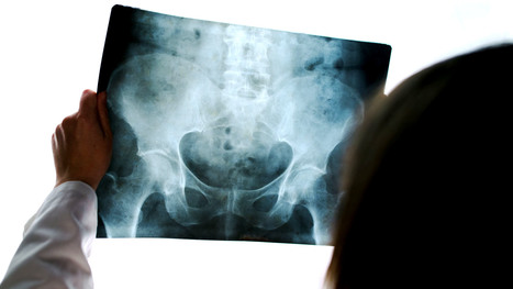
Pelvic Fractures Can Be Stable & Unstable - Unstable Fx: a result of high energy trauma with >50% d/t MVA
- 20% closed Fx and 50% of open Fx result in mortality
- Mortality is associated with vascular and internal organs injuries
- Vascular injury: 20% arterial 80% venous
- Chronic morbidity/disability and prolonged pain
- Unstable Fx are rarely seen in the outpatient setting and typically and present to the ED
- Stable pelvic Fx are usually caused by muscles/tendons avulsions and more often seen in pediatric cases
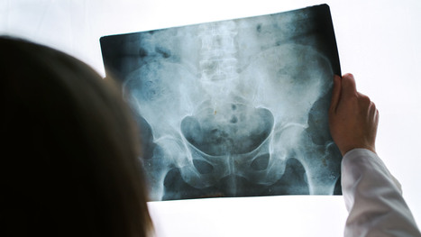
Understanding Pelvic Anatomy Is The Key To Successful Imaging Dx - The bony pelvis is a continuous ring of bone held by strong ligaments
- During significant impact, pelvic fractures may occur in more than one location because forces applied to one region of the ring will also correspond to injury on the other, usually the opposite side of the ring (above image)
- Thus the majority of unstable pelvic Fx will typically demonstrate more than one break
- Pelvic is seen as a ring of bone connected by some of the strongest ligaments in the body
- The pelvic ring comprises 2-semirings: anterior to the acetabulum and posterior to the acetabulum
- The bony pelvis is in close proximity to major vessels carrying a greater chance of vascular injury
- Anatomical Differences of The Female and Male Pelvis
Post-Traumatic Pelvic Views May Vary and Include: - Standard AP Pelvis (above images)
- Judet views evaluating the acetabulo-pelvic region
- Inlet/Outlet views helping with the symphysis and SIJ regions
- Rad survey of the pelvis should include evaluation of the continuity of pelvic rings:
- Inlet/outlet, obturator rings (above the first image)
- Symphysis pubis and SIJ for diastasis and post-trauma separation (above the second image)
- Lumbosacral spine and hips should also be carefully examined
- Pelvic inlet (above top left) and Outlet (above bottom left)
- Judet views: left and right posterior oblique views
Additional Survey: - Iliopectineal, ilioischial, Shenton and Sacral arcuate lines will help detection of sacral, acetabular and hip fracture/dislocations
Stable Pelvic Fractures aka Avulsion Injury - Appreciating anatomical sites of pelvic origin/insertion of different muscles will help Dx of pelvic avulsion Fx
- Avulsion Fx of the AllS (origin of the direct head of Rectus femoris M)
- Pelvic avulsions occur by sudden eccentric contraction especially during kicking or jumping
- Imaging: x-radiography will suffice
- Clinically: sudden snap or pop followed by local pain. Pt can weight bear
- Care: non-operative with rest for 4-weeks. Non-union is rare. No major complications
- DDx: key rad DDx feature is not to mistake an avulsion from an aggressive pediatric bone tumor-like osteosarcoma that may show some exuberant new bone formation d/t healing and bone callus
Commonly Encountered Unstable Pelvic Fractures - Malgaigne Fx: d/t vertical shear injury to the ipsilateral pelvis
- Rad Dx: ipsilateral superior and inferior pubic rami Fx (anterior ring) with ipsilateral SIJ separation/Fx of the sacrum and adjacent ilium (posterior ring). Symphysis pubis diastasis can be seen. An additional clue is an avulsion of L4 and/or L5 TP that often signifies serious pelvic injury
- Clinically: marked leg shortening, shock, inability to weight bear.
- Damage to Superior Gluteal Artery can occur
- Imaging: x-radiography followed by CT scanning w/o and with IV contrast esp. if visceral injury present
- Care: surgical in most cases d/t significant instability. ORIF. Hemostasis, Pelvic stabilization
- Prognosis: depends on the complexity, rate of visceral complications and stability. 10% Superior glut artery bleed requiring rapid hemostasis
Open Book Pelvis (major instability) - Mechanism: AP compression of different force magnitude (picture depiction)
- Rad Dx: diastasis of symphysis pubis with diastasis of SIJ with and w/o adjacent Fx of the ala
- Imaging steps: x-radiographic, CT scanning with and w/o contrast for vascular injury, cystography for acute urinary bladder rupture
- Immediate and delayed complications may occur: vascular injury, urethral/bladder injury
Straddle Injury: Unstable Fx - Mechanism: direct impact/collision
- High risk of urinary bladder/urethral injury
- Imaging: bilateral superior and inferior pubic rami Fx with or w/o diastasis and Fx of SIJ
- CT with and w/o contrast for vascular injury
- Cystourethrogram additionally evaluates a urogenital injury
- Complications: urethral strictures, bleeding, bladder rupture
- Note: Straddle Fx with right SIJ separation
Hip Fractures (Femoral Neck) - Common injury
- Occurs in:
- 1) Young adults due to high energy trauma
- 2) Osteoporotic patients with low impact, trivial or no trauma (i.e., insufficiency Fx)
- X-radiography is crucial to early Dx and prevention of complications which include:
- Dx: intra-capsular vs. extra-capsular Fx
- Ischemic osteonecrosis aka avascular necrosis (AVN) of the femoral head and rapid disabling DJD
- Epidemiology: USA has some of the highest rates of OSP hip Fx worldwide. Highest healthcare cost Fx to treat overall
- Women>men, Caucasians>African-Americans
- 25-30% mortality within the 1st year. Mortality depends on co-morbidities and stat of activity prior Fx
- Pathophys: the femoral neck is intra-capsular and transmits arterial flow to the head. The neck is uncovered by the periosteum and unable to develop a good callus. The neck transmits maximum tensile forces through the proximal femur and prone to Fx and non-union
Acute Pelvis & Hip Trauma
|


Scar tissue is part of the healing process but the tissue build-up can restrict mobility, range of motion, and lack of flexibility. For answers to any questions you may have please call Dr. Jimenez at 915-850-0900 or 915-412-6677